Medizinische Mikrobiologie & Krankenhaushygiene


Arbeitsgruppe Dr. Tobias Geiger

Die Nachwuchsgruppe der AG Geiger ist seit Ende 2019 am Max von Pettenkofer Institut tätig. Das wissenschaftliche Interesse liegt im Verständnis molekularer Mechanismen, die der Pathogenität von gastrointestinalen und systemischen Pathogenen zu Grunde liegen. Die Interessen umfassen dabei im Besonderen die Charakterisierung von Sekretionssystemen, Sekretionsmechanismen, immunmodulatorischen Effektorproteinen sowie Virulenzfaktoren, die für die Wirt/Pathogen Interaktion wichtig sind. Unser Focus liegt hier bei typhoidalen Salmonellen (Serovare Typhi und Paratyphi A), fakultativ intrazelluläre Pathogene, die ursächlich für das Typhus Fieber im Menschen verantwortlich sind. Diese Serovare infizieren ausschließlich Menschen und können neben der Pathogenese langfristig im menschlichen Körper persistieren. Unsere Studien sollen zu neuen Ansätzen in der Prävention, Diagnose und Therapie von Typhus Infektionserkrankungen führen.

Zur Arbeitsgruppe

Leitung

Dr. rer. nat. Tobias Geiger

The studies of biology at the University of Tübingen with focus on microbiology, biochemistry and genetics drew my interests towards intracellular pathogens and their interactions with the host. For my PhD, I joined the lab of Prof. Dr. Christiane Wolz at the Institute of Medical Microbiology and Hygiene in Tübingen, where I focused on the pathogenicity and intracellular behavior of Staphylococcus aureus, including methicillin-resistant S. aureus (MRSA). After my PhD, I extended my stay in Tübingen working as a Postdoctoral Associate on S. aureus. In 2014, I received a Postdoctoral Fellowship to join the lab of Prof. Dr. Jorge E. Galán at the Yale University in New Haven, CT USA. Here, I studied intracellular Salmonella typhi and the secretion of typhoid toxin as a Postdoc and later as an Associate Research Scientist. Since the end of 2019, I am an independent Junior Research Group leader at the Max von Pettenkofer Institute of the Ludwig Maximilian University (LMU) in Munich.

Dr. rer. nat. Tobias Geiger

AG-Mitglieder

Aktuelle Gruppenmitglieder

Dr. Tobias Geiger, (group leader)
E-Mail: geiger@mvp.lmu.de
Phone: +49 89 2180-72845

Lena Krone, M.Sc. (doctoral researcher)
E-Mail: krone@mvp.lmu.de
Phone: +49 89 2180-72846

Srujita Mahankali, M.Sc.(doctoral researcher)
E-Mail: mahankali@mvp.lmu.de
Phone: +49 89 2180-72846

Forschung

A Novel Protein Secretion System for Virulence Factors

Bacterial pathogens have evolved sophisticated protein secretion systems that perform numerous physiological functions essential for cell propagation and fitness within a specific ecological niche. These secretion systems play a pivotal role in the pathogenicity of bacterial pathogens by facilitating attachment to various surfaces, defense against host antimicrobial responses, delivery of virulence factors and effector proteins to the extracellular environment or into eukaryotic cells, nutrient acquisition and communication with the surrounding environment.

Protein transport across the bacterial envelope is particularly challenging in Gram-negative bacteria, as proteins must move through at least three barriers: the inner membrane (IM), the peptidoglycan (PG) layer and the outer membrane (OM). Therefore, Gram-negative bacteria have evolved multiple secretion mechanisms of varying complexities to transport proteins across the bacterial envelope. While some complex protein secretion machines with designated cell envelope-spanning structures move their substrates through all the layers of the bacterial envelope in one step (Type 1, Type 3, Type 4 and Type 6 secretion systems), others operate by engaging the substrates in the bacterial periplasm after their translocation by dedicated IM proteins (Type 2 secretion system).

Our work on typhoid toxin in Salmonella enterica serovar Typhi (S. typhi) revealed a protein secretion system, that differs significantly from the well-described secretion systems of the various types mentioned before. In this secretion system, a specialized N-acetyl-β-D muramidase named TtsA (typhoid toxin secretion protein A) represents the essential component facilitating the secretion of typhoid toxin, a major virulence factor of S. typhi.

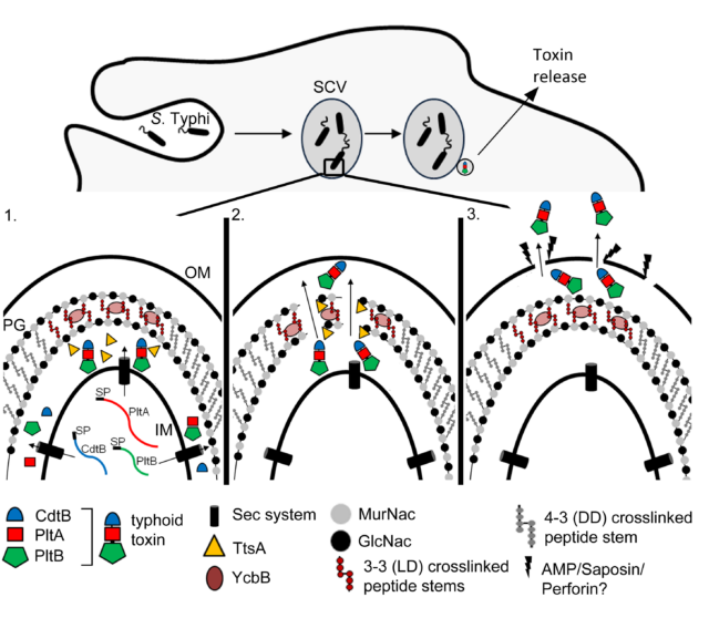
The current model for typhoid toxin secretion is a 4-steps release mechanism. First, within the SCV the peptidoglycan (PG) of S. typhi is modified by a periplasmic LD-transpeptidase YcbB, that introduces 3-3 crosslinks into the PG layer.  In step 2, TtsA is translocated to the periplasm, where it hydrolyzes the PG layer with its specific activity towards 3-3 crosslink modified peptidoglycan. In step 3, the periplasmic and fully assembled typhoid toxin crosses the TtsA-edited PG layer and accumulates beneath the outer membrane (OM). In a final step, minor disruptions of the OM inflicted by membrane active agents such as antimicrobial peptides, commonly found within the SCV, finally release the toxin to the extracellular space (Geiger et al., 2018).

Genomic analyses have shown that homologous muramidases or peptidoglycan hydrolases encoded in close proximity to toxins or other virulence factors exist in many bacterial genomes. These analyses, which also includes studies in bacterial pathogens such as Serratia and Clostridia have shown that the peptidoglycan hydrolase-dependent protein secretion system is a conserved mechanism for protein secretion in many bacteria.

Albeit progress has been made to shed light on this novel protein secretion mechanism, many aspects still remain unclear and are currently under investigation in the lab.

Current model for the typhoid toxin secretion mechanism

Specific virulence factors of human-adapted Salmonella enterica serovar Typhi and serovar Paratyphi A

The virulence of S. typhi differs significantly from closely related, broad host range serovars of Salmonella enterica, such as Salmonella enterica serovar Typhimurium (S. typhimurium), which are a common source of food poisoning. These “generalists” serovars typically cause short-term infections that remain confined to the gastrointestinal tract in healthy humans. By contrast, typhoidal serovars such as S. typhi lead to systemic infections and, for a subset of those who suffer the disease, they cause life-long chronic infections. Long-term carriers shed high levels of S. typhi asymptomatically and are thought to be crucial for the transmission of typhoid fever since humans are the only known reservoir for S. typhi. In recent years, multiple antibiotic resistant strains have begun to emerge, raising the alarming prospect of untreatable typhoid fever. Therefore, this situation desperately requires a greater understanding of the pathogenicity of typhoidal Salmonella. Little is known about the specific factors that confer on this pathogen its unique pathogenicity. It is believed that genome reduction and the acquisition of unique genes are responsible for the remarkable differences in the disease presentation and host range. The genome of S. typhi contains an unusually large number of pseudogenes compared to nucleotide sequences of other S. enterica serovars. This observation has led to the hypothesis that the reduction of its genome may have contributed to S. Typhi´s exclusive restriction to the human host. The genome sequencing of S. typhi also revealed many genes unique to this serovar. Some of these unique genes encode for putative virulence factors, which may contribute to its distinctive virulence properties.

Working with S. typhi for many years, we found unique virulence factors, that are responsible for the increased virulence of S. typhi. These factors are currently under investigation regarding their impact on immune cell evasion and their contributions leading to a systemic infection of humans by S. typhi.


Publikationen

Top 10 Publikationen

Krone L, Mahankali S, Geiger T. Cytolysin A is an intracellularly induced and secreted cytotoxin of typhoidal Salmonella. Nat Commun. 2024 Sep 28;15(1):8414. doi: 10.1038/s41467-024-52745-0. PMID: 39341826
Krone L, Faass L, Hauke M, Josenhans C, Geiger T. Chitinase A, a tightly regulated virulence factor of Salmonella enterica serovar Typhimurium, is actively secreted by a Type 10 Secretion System. PLoS Pathog. 2023 Apr 5;19(4):e1011306. doi: 10.1371/journal.ppat.1011306. eCollection 2023 Apr. PMID: 37018381
Geiger T, Lara-Tejero M, Xiong Y, Galán JE. Mechanisms of substrate recognition by a typhoid toxin secretion-associated muramidase. Elife. 2020;9:e53473. Published 2020 Jan 20. doi:10.7554/eLife.53473.
Geiger T , Pazos M , Lara-Tejero M , Vollmer W, Galán JE. Peptidoglycan editing by a specific LD-transpeptidase controls the muramidase-dependent secretion of typhoid toxin. Nature Microbiology, 2018 Nov;3(11):1243-1254. doi:10.1038/s41564-018-0248-x
Gratani FL, Horvatek P, Geiger T, Borisova M, Mayer C, Grin I, Wagner S, Steinchen W, Bange G, Velic A, Maček B, Wolz C. Regulation of the opposing (p)ppGpp synthetase and hydrolase activities in a bifunctional RelA/SpoT homologue from Staphylococcus aureus. PLOS Genetics. 2018 Jul 9;14(7)
Münzenmayer L, Geiger T, Daiber E, Schulte B, Autenrieth SE, Fraunholz M, Wolz C. Influence of Sae-regulated and Agr-regulated factors on the escape of Staphylococcus aureus from human macrophages. Cellular Microbiology. 2016 Aug;18(8)
George SE, Nguyen T, Geiger T, Weidenmaier C, Lee J, Liese J, Wolz C. Phenotypic heterogeneity and temporal expression of the capsular polysaccharide in Staphylococcus aureus. Molecular Microbiology. 2015 Dec;98(6):1073-88.
Grosz M, Kolter J, Paprotka K, Winkler AC, Schäfer D, Chatterjee SS, Geiger T, Wolz C, Ohlsen K, Otto M, Rudel T, Sinha B, Fraunholz M. Cytoplasmic replication of Staphylococcus aureus upon phagosomal escape triggered by phenol-soluble modulin α. Cellular Microbiology. 2014 Apr;16(4).
Geiger T, Kästle B, Gratani FL, Goerke C, Wolz C. Two Small (p)ppGpp Synthases in Staphylococcus aureus Mediate Tolerance against Cell Envelope Stress Conditions. Journal of Bacteriology. 2014 Feb;196(4):894-902.
Geiger T, Francois P, Liebere M, Fraunholz M, Goerke C, Krismer B, Schrenzel J, Lalk M, Wolz C. The Stringent Response of Staphylococcus aureus and its impact on survival after phagocytosis through the induction of intracellular PSMs expression. PLOS Pathogens. 2012 Nov; 8(11).