Virologie


Arbeitsgruppe PD Dr. rer. nat. Dr. habil. med. Albrecht von Brunn

Die AG von Brunn untersucht und identifiziert Protein-Protein Interaktionen zoonotischer Viren sowohl auf intraviraler als auch auf Virus-Wirtsebene. Ziel ist die Entwicklung von Breitspektrum Virostatika über die Identifizierung fundamentaler Prozesse und Signalwege der viralen Replikation in infizierten Wirtszellen. Hierbei liegt das Hauptaugenmerk auf Coronaviren einschließlich mild pathogener Erkältungsviren und hoch pathogener SARS-CoV/SARS-CoV-2 und MERS-CoV, sowie auf Zika Virus. Die Strategie trägt zum prä-pandemischen und pandemischen Vorbereitetsein gegenüber neu auftretenden viralen Infektionskrankheiten bei.

Zur Arbeitsgruppe

Leitung

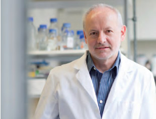
PD Dr. rer. nat. Dr. habil. med. Albrecht von Brunn

Albrecht von Brunn, PhD, is principal investigator in Virology at the Max von Pettenkofer Institute at LMU Munich. Albrecht von Brunn grew up in Northern Baden-Württemberg, where he obtained primary and secondary school education. Here he also fulfilled his compulsory military service. From the Albert-Ludwigs-Universität Freiburg, Germany he received his “Vordiplom” degree in Biology.  He received graduate student scholarships from the University of Texas at Austin and from the Marine Biological Laboratory, Woods Hole, MA, USA, and after two years of graduate studies at UT Austin he graduated with a Masters degree. He was a visiting scientist at the European Molecular Biology Laboratory (EMBL) Heidelberg. The doctoral thesis he performed at the Center for Molecular Biology (ZmBH)/Ruprecht-Karls-Universität Heidelberg under the supervision of Prof. Herrmann Bujard and Prof. Heinz Schaller. Upon completion of his dissertation in Molecular Biology he assumed a principal investigator position at the Max von Pettenkofer Institute in Munich, where he also received his “Habilitation” in Experimental Virology.

AG-Mitglieder

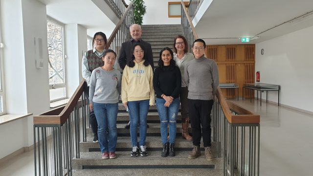
Aktuelle Gruppenmitglieder

von Brunn, Albrecht, PD Dr. rer. nat. Dr. habil. med., principal investigator
E-Mail: vonbrunn@mvp.lmu.de
Phone: +49 89 2180-72839

Baral, Priya, cand. med., MD student (FöFoLe programme)
E-Mail: Baral@mvp.lmu.de
Phone: +49 89 2180-72891

Li, Pengyan, CSC scholar, doctoral student
E-Mail: Pengyuan.Li@mvp.lmu.de
Phone: +49 89 2180-72891

Ma-Lauer, Yue, Dr. rer. nat., post-graduate researcher
E-Mail: Ma_Lauer@mvp.lmu.de
Phone: Phone: +49 89 2180-72889

Konstantin Pusl, MD

Ru, Yi, doctoral student
E-Mail: Ru@mvp.lmu.de
Phone: +49 89 2180-72891

von Brunn, Brigitte, MTLA
E-Mail: vonbrunn_b@mvp.lmu.de
Phone: +49 89 21807-2891

Xiang, Chengyu, doctoral student
E-Mail: Xiang@mvp.lmu.de
Phone: +49 89 2180-72891


Ehemalige Gruppenmitglieder

  • Emilia Berthold, (MD predoctoral student, together with PD Dr.
  • Claudia-Staab-Weijnitz)
  • Javier Carbajo, (technician)
  • Dev Raj Bairad, (predoctoral student)
  • Jürgen Huber, ( MD predoctoral student: Dr. med.)
  • Marisa Kurz, (MSc student: MA Biochemistry)
  • Peter Mayerhofer, (MSc student: MA Biochemistry)
  • Su Mingxia, (postdoctoral researcher)
  • Julia Schöpf, (predoctoral student: Dr. rer. Nat.)
  • Sophia Schwoier, (BSc internship student, studentische Hilfskraft)
  • Nicole Senninger, (technician)
  • Wulf Sienel, (MD student: Dr. med.)
  • Thorsten Stellberger, (predoctoral student, postdoctoral researcher)
  • Carola Teepe, (FoeFoLe MD student: Dr. med.)
  • Nadja Wermke, (technician)
  • Harald Wizemann, (predoctoral student: Dr. rer. Nat.)
  • Zheng Yu, (postdoctoral researcher)
  • Sebastian Schwinghammer, (MD, Dr. med.)

Internships/studentische Hilfskräfte

  • Mustafa Abdellatif, (Mol. & Cell. Biology internship student, studentische Hilfskraft)
  • Kemal Baskaya, (BSc internship student, BSc student: BSc, studentische Hilfskraft)
  • Arthur Brommer, (MA Human Biology internship student, studentische Hilfskraft)
  • Sarah Christ, (MSc internship student)
  • Manuela Decker, (MA internship student Nutritinal Sciences TUM)
  • Simon Ebert, (MA Biochemistry internship student)
  • Allison Maher, (MA internship student, University of Oxford)
  • Thomas Höltke, (MA Biochemistry internship student)
  • Larissa Knüppel, (BA Biochemistry internship student)
  • Jennifer Mittermaier, (MA Human Biology internship student)
  • Baopeng Sha, (MA Biochemistry internship student)
  • Sophia Schwojer, (MA Biochemistry internship student, studentische Hilfskraft)
©AvB
©AvB

Forschung

Coronaviruses (CoVs, family Coronaviridae, subfamily Orthocoronavirinae) are important human and animal pathogens that induce fatal respiratory, gastrointestinal and neurological disease. Seven distinct CoVs (HCoV-NL63, HCoV-HKU-1, HCoV-OC43, HCoV-229E, SARS-CoV, HCoV-MERS, SARS-CoV-2) cause respiratory tract illnesses in humans, ranging from mild common cold infections in immune-competent individuals to deadly virus-associated pneumonia, organ failure etc. At least seven different animal CoVs cause economically significant epizootics in livestock, and some cause deadly disease in companion animals. Some CoV have zoonotic potential and are considered as emerging viruses. The recent CoV outbreaks and current SARS-CoV-2/COVID-19 pandemic demonstrate the necessity to develop highly effective, broadly-acting compounds against zoonotic viruses.

The laboratory studies the interplay between viral and host cell proteins of mainly coronaviruses in order to identify and understand cellular determinants and pathways responsible for different pathogenicity. Goal is to identify common targets for the development of broadly acting antivirals.

Identification of protein-protein interactions at intraviral and virus-host levels by high throughput Yeast-2-Hybrid (Y2H) screening technologies

CoVs are enveloped viruses carrying the largest known single-stranded RNA genomes (25–32 kb) with positive-sense orientation. The first two thirds of the genomes encode two polyproteins ORF1a/ORF1b, which are processed by viral proteases into 16 non-structural proteins with various enzymatic functions required for genome replication. The last third of the genome contains four structural proteins S, E, M, N and – depending on the virus – various accessory genes. To understand different pathogenicity of the CoV family members it is essential to gain knowledge on the function of the individual viral proteins, their interaction with cellular proteins and the consequences of these interactions on cellular signaling pathways.

Using high-throughput Y2H screening technologies, our lab was one of the first to study protein/protein interactions (PPIs) of individual SARS-CoV proteins at intra-viral, matrix-based (screening all viral proteins against each other) as well as virus-host levels (screening viral ORFs against human cDNA libraries)/(Pfefferle et al., 2011).

We have constructed orfeomes of SARS-CoV, SARS-CoV-2, MERS-CoV, HCoV-NL63 by PCR amplification of all ORFs and a number of sub-fragments lacking transmembrane regions of the respective CoV from cloned viral DNA. Fragments are cloned into GATEWAY™- compatible pDONR vectors. From there they can easily be shuttled into pro- and/or eukaryotic (Y2H, mammalian) destination expression vectors. Mammalian gene products interacting with viral proteins are identified by screening individual CoV ORFs against human, yeast-expressed cDNA- encoded genes. Positive hits are confirmed in mammalian cells by a variety of techniques including split YFP, LUMIER- and CoIP assays, and in collaboration with several labs Bimolecular Fluorescence Complementation, Fluorescent Three Hybrid, Mass Spec, X-ray crystallography (Ma-Lauer et al., PNAS 2016; Lei et al., 2021). We have available a battery of expression vectors with various N- and C- terminal fusion tags (e.g. HA, c-myc, HIS, GFP, RFP, split YFP).

We anticipate the identification of cellular molecules and pathways explaining different pathogenicities of the viruses, and serving as common antiviral targets.

The power of our virus-host Y2H protein-protein interaction screening approaches for successful identification of signaling pathways and antiviral targets is illustrated e.g. by the identification of:

a) immunophilins (cyclophilins, FK506-binding proteins [FKBPs]) as essential for correct folding of viral proteins, and inhibitors with broad-spectrum antiviral potential,

b) E3- dependent ubiquitinating and proteasomal degradation of the anti-virally active p53 tumor protein and

c) influence of PAIP1 (Poly(A) Binding Protein Interacting Protein 1) on mRNA translation in virus-infected host cells.

Details:

a) Involvement of immunophilins in CoV replication and broad-spectrum inhibition

Cyclophilins and FKBPs represent large families of peptidyl-prolyl cis/trans isomerases (PPIases) with chaperone-like activities thus excerting important functions on folding, maturation and trafficking of proteins within the eukaryotic cell. Cyclosporin A (CsA) acts as a tight-binding, reversible, competitive inhibitor of the PPIase activity. Cyclophilins directly interact with cellular proteins, and in some cases also with viral proteins thus granting replication sensitivity to CsA (e.g. HIV, HCV).

Inhibiting molecules CsA and FK506 (Tacrolimus) modulate the calcineurin A (CnA)/NFAT (Nuclear Factor of Activated T cells) pathway, which plays an important role in immune-cell activation. These compounds are used to suppress the immune system in transplant patients. Both, protein folding and immunosuppressive activites can be separated by chemically modifying side chains of e.g. CsA, resulting in non-immunosuppressive, enzymatic function- inhibiting derivatives like Alisporivir (Debio 025).

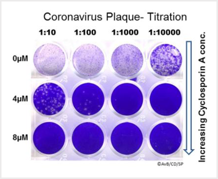
Using Y2H screening methods we identified Non-structural protein Nsp1 and other proteins of various coronaviruses, including SARS-CoV and SARS-CoV-2, to interact with immunophilins. For CoVs we and our collaborators were the first to demonstrate that inhibition of cyclophilins by CsA blocks the replication of representatives of the Orthocoronavirinae subfamily, including human SARS-CoV (Figure 1), HCoV-229E and -NL63, feline CoV, Mouse Hepatitis Virus (MHV) as well as avian infectious bronchitis virus (Pfefferle et al., 2011). Similar results were found using a number of different non-immunosuppressive derivatives including Alisporivir (Carbajo-Lozoya et al., 2014; Ma-Lauer et al., 2020). Latter compounds are especially interesting as they do not suppress the host immune system but retain their antiviral activity. Similarly, we have shown that replication of human CoVs SARS-CoV, HCoV-NL63, and HCoV-229E is inhibited by the drug FK506 (Tacrolimus; Carbajo-Lozoya et al., 2012).

The results indicate that these Host-Targeting-Agents (HTAs) carry the potential to serve as broad-range inhibitors applicable against emerging CoVs as well as ubiquitous pathogens of humans and livestock. Investigations on mechanistic principles are carried out further.

Figure 1: Plaque Titration of a CoV (here SARS-CoV) in the presence of various concentrations of cyclosporin A. The virus causes plaques in a monolayer of CaCo2 cells indicating the number of infectious virus particles. At increasing concentrations of CsA virus particles decrease. They are not detectable any more at optimal inhibitor concentration.

b) Influence of tumor suppressor protein p53 on SARS-CoV replication

Our protein-protein interactions screenings at virus-host levels revealed an indirect link between the SARS-CoV SUD (SARS Unique Domain) and PLPro domains of non-structural protein nsp3 and p53: we found that both CoV domains bind to and stabilize the host E3 ubiquitin ligase RCHY1 (Ring Finger And CHY Zinc Finger Domain Containing 1). This enzymes marks target proteins by ubiquitination (addition of small ubiquitin protein molecules) thus changing their properties. One consequence is the degradation of the ubiquitinated proteins in the host-cell proteasome complex. p53 is one of the targets of RCHY1. p53 regulates a number of target genes that mediate tumor suppression. For some viruses it deploys anti-viral activity. We tested a possible functional link between p53 and SARS-CoV replication by infection cells lacking p53. Indeed, we found that in p53-negative cells replication of SARS-CoV was several orders of magnitude more efficient than in p53-expressing cells (Ma-Lauer et al., 2016). As p53 regulates genes involved in the non-specific antiviral defense system (innate immunity) we hypothesize that the degradation of p53 is actively promoted by viral domains stabilizing the RCHY1 enzyme.

c) Influence of Poly(A) Binding Protein Interacting Protein 1 (PAIP1) on SARS-CoV/-2 mRNA translation

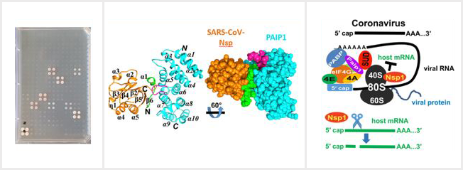
By Y2H virus-host protein-protein interaction screening we further identified the interaction of SARS-CoV Nsp3/SUD with human PAIP1, a stimulator of protein translation. In collaboration with the Rolf Hilgenfeld laboratory, University of Lübeck, we subsequently validated SARS-CoV SUD:PAIP1 interaction by size exclusion chromatography, split-yellow fluorescent protein and co-immunoprecipitation assays. Such interaction we also confirmed between the corresponding domain of SARS-CoV-2 and PAIP1. The three-dimensional structure of the N-terminal domain of SARS-CoV SUD (“macrodomain II”, Mac2) in complex with the middle domain of PAIP1, determined by X-ray crystallography and small angle X-ray scattering, provides insights into the structural determinants of the complex formation (Figure 2). In cellulo, SUD enhances synthesis of viral but not host proteins via binding to PAIP1 in pBAC-SARS-CoV replicon-transfected cells. We propose a possible mechanism for stimulation of viral translation by Nsp3-SUD of SARS-CoV and SARS-CoV-2 (Lei et al., 2021).

Figure 2: From Y2H protein-protein interaction screening of SARS-CoV/-2 ORFs with host cell genes to crystal structure and translation regulation analysis (Lei et al., EMBO J. 2021, 40(11):e102277, open access)

Future directions

Conventionally, antiviral drugs are directed against viral enzymes/proteins. The selective pressure to mutate e.g. active centers of replicative enzymes or proteases is very high. Our philosophy is that this is not the case for cellular protein targets because the respective genes of the infected eukaryotic cell do not „mutate away” easily, thus changing amino acids and protein structures upon selective drug pressure. The barriers for developing host-factor resistance is expected to be much higher. Therefore, we are following up various projects characterizing different protein-protein interactions of viral and cellular counterparts identified by our various screening projects.

Publikationen

Top 10 Publikationen

Ma-Lauer Y, Li P, Niemeyer D, Richter A, Pusl K, von Brunn B, Ru Y, Xiang C, Schwinghammer S, Liu J, Baral P, Berthold EJ, Qiu H, Roy A, Kremmer E, Flaswinkel H, Drosten C, Jin Z, von Brunn A. (2024). Oxysterole-binding protein targeted by SARS-CoV-2 viral proteins regulates coronavirus replication. Front. Cell. Infect. Microbiol. 2024; 14:1383917. https://doi.org/10.3389/fcimb.2024.1383917
Berthold EJ, Ma-Lauer Y, Chakraborty A, von Brunn B, Hilgendorff A, Hatz R, Behr J, Hausch F, Staab-Weijnitz CA*, von Brunn A* (2022). Effects of immunophilin inhibitors and nonimmunosuppressive analogs on coronavirus replication in human infection models. Front. Cell. Infect. Microbiol. 2022, 12:958634. https://doi.org/10.3389/fcimb.2022.958634
Lei J†, Ma-Lauer Y†, Han Y, Thoms M, Buschauer R, Jores J, Thiel V, Beckmann R, Deng W, Leonhardt H, Hilgenfeld R*, von Brunn A* (2021). The SARS-unique domain (SUD) of SARS-CoV and SARS-CoV-2 interacts with human PAIP1 to enhance viral RNA translation. The EMBO Journal 1;40(11):e102277. https://doi.org/10.15252/embj.2019102277
Poulsen, NN, von Brunn, A, Hornum, M and Blomberg, JM (2020) Cyclosporine and COVID-19: Risk or Favorable? Am J Transplant. (2020). 20:2975-2982. https://doi.org/10.1111/ajt.16250
Ma-Lauer Y, Zheng, Y Malešević M, von Brunn B, Fischer F, von Brunn A* (2020). Influences of cyclosporin A and non-immunosuppressive derivatives on cellular cyclophilins and viral nucleocapsid protein during human coronavirus 229E replication. Antiviral Research 173, 104620. https://doi.org/10.1016/j.antiviral.2019.104620
Ma-Lauer Y, Carbajo-Lozoya J, Hein M, Müller MA, Deng W, Lei J, Meyer B, Kusov Y, von Brunn B, Bairad DR, Hünten S, Drosten C, Hermeking H, Leonhardt, H, Mann M, Hilgenfeld R, von Brunn, A* (2016). p53 down-regulates SARS-Coronavirus replication and is targeted by the SARS-Unique Domain and PLpro via E3 ubiquitin ligase RCHY1 Proc Natl Acad Sci U S A, 113(35): E5192-201. www.pnas.org/cgi/doi/10.1073/pnas.1603435113
Carbajo-Lozoya, J., Ma-Lauer, Y., Malešević, M., Theuerkorn, M., Kahlert, V., Prell, E., von Brunn, B., Muth, D., Baumert, T.F., Drosten, C., Fischer, G., and von Brunn, A* (2014). Human coronavirus NL63 replication is cyclophilin A-dependent and inhibited by non-immunosuppressive cyclosporine A-derivatives including Alisporivir. Virus Research 184, 44-53. https://doi.org/10.1016/j.virusres.2014.02.010
Carbajo-Lozoya, J., Müller, M.A., Kallies, S., Thiel, V., Drosten, C. and von Brunn, A* (2012). Replication of human coronaviruses SARS-CoV, HCoV-NL63 and HCoV-229E is inhibited by the drug FK506. Virus Research 165, 112–117. https://doi.org/10.1016/j.virusres.2012.02.002
Pfefferle S, Schöpf J, Kögl M, Friedel C, Müller MA, Carbajo-Lozoya J, Stellberger T, von Dall'Armi E, Herzog P, Kallies S, Niemeyer D, Ditt V, Kuri T, Züst R, Pumpor K, Hilgenfeld R, Schwarz F, Zimmer, R., Steffen I, Weber F, Thiel V, Herrler G, Thiel H-J, Schwegmann-Weßels, C., Pöhlmann S, Haas J, Drosten C, von Brunn, A* (2011). The SARS-Coronavirus-Host Interactome: Identification of Cyclophilins as Target for Pan-Coronavirus Inhibitors. PLoS Pathogens 7(10): e1002331. https://doi.org/10.1371/journal.ppat.1002331
von Brunn A*, Teepe C, Simpson JC, Pepperkok R, Friedel C., Zimmer R, Roberts R, Baric R, Haas, J* (2007). Analysis of Intraviral Protein-Protein Interactions of the SARS Coronavirus ORFeome. PLoS ONE 23;2:e459. https://doi.org/10.1371/journal.pone.0000459

Auszeichnungen

Auszeichnungen

  • Graduate Student Scholarship, The University of Texas at Austin, Austin, TX, USA
  • Marine Biological Laboratory, Woods Hole, MA, USA, Walter E. Garrey Scholarship
  • Young Investigator Research Award of the „Gesellschaft zur Förderung der Molekularbiologie Heidelberg e.V. (GFM)“